Some recent papers:
- Huang J., Ruzhansky M., Feng H., Zheng L., Huang X., Wang H., Feature extraction for license plate location based on L0-norm smoothing. Open Comput. Sci. 2019; 9:28-135. link (open access)
- Mamaeva S.N., Kononova I.V., Ruzhansky M., Nikiforov P.V., Nikolaevа N.A., Pavlov A.N., Fedorova N.F., Huang J., Semenova M.N., Barashkova D.V., Frolova L.S., Maksimov G.V., Using Scanning Electron Microscopy and Atomic Force Microscopy to Study the Formation of Nanoparticles on Red Blood Cell Surface in Cervical Cancer Patients, International Journal of Biomedicine, 10(1): 70-75, 2020. link (open access)
- Huang J., Wang H., Wang X., Ruzhansky M., Semi-sparsity for smoothing filters. IEEE Transactions on Image Processing, vol. 32, pp. 1627-1639, 2023. doi, arxiv
- Huang J., Ruzhansky M., Zhang Q., Wang H., Intrinsic image transfer for illumination manipulation. IEEE Transactions on Pattern Analysis and Machine Intelligence, vol. 45, no. 6, pp. 7444-7456, 2023. doi, arxiv
- Wang X., Huang J., Chatzakou M., Nomm S., Valla E., Medijainen K., Taba P., Toomela A., Ruzhansky M., Comparison of one- two- and three-dimensional CNN models for drawing-test-based diagnostics of the Parkinson’s disease. Biomedical Signal Processing and Control, 87 (2024) 105436. doi, arxiv
- Huang J., Wang H., Ruzhansky M., Semi-sparsity on piecewise constant function spaces for triangular mesh denoising. arxiv
- Huang J., Wang H., Ruzhansky M., Semi-sparsity priors for image structure analysis and extraction. arxiv
- Wang X., Huang J., Chatzakou M., Medijainen K., Taba P., Toomela A., Nomm S., Ruzhansky M., A Light-weight CNN Model for Efficient Parkinson’s Disease Diagnostics. arxiv
- Huang J., Wang H., Weiermann A., Ruzhansky M., Optimal image transport on sparse dictionaries. arxiv
- Wang X., Huang J., Nomm S., Chatzakou M., Medijainen K., Toomela A., Ruzhansky M., LSTM-CNN: An efficient diagnostic network for Parkinson’s disease utilizing dynamic handwriting analysis. arxiv
Some modelling of COVID-19
The effectiveness analysis is carried our for the lockdown in Belgium and of its duration, as well as of several phases of its relaxation. The comparative projection is made of different scenarios depending on the strength of confinement measures and their implementation.
Ruzhansky M., Tokmagambetov N., Torebek B., A projection model of COVID-19 pandemic for Belgium. medRxiv, paper
Agarwal P., Nieto J., Ruzhansky M., Torres D. (Eds.) Analysis of Infectious Disease Problems (Covid-19) and Their Global Impact, Infosys Science Foundation Series in Mathematical Sciences, Springer, 2021. link, doi
Imaging analysis
(joint with Beijing University of Aeronautics and Astronautics (BUAA), China)
Real-time image/video enhancement for liver endoscopy
This is research in progress. Some related papers can be seen:
- Huang J., Ruzhansky M., Feng H., Zheng L., Huang X., Wang H., Feature extraction for license plate location based on L0-norm smoothing. Open Comput. Sci. 2019; 9:28-135. link (open access)
- Huang, J., Ruzhansky M., Wang, H. (2020). Weakly supervised learning photo enhancer with inexact training pairs.
- Nikiforov, P. V., Nikolaevа, N. A., Pavlov, A. N., Fedorova, N. F., Huang, J., Semenova, M. N., … & Frolova, L. S. Using Scanning Electron Microscopy and Atomic Force Microscopy to Study the Formation of Nanoparticles on Red Blood Cell Surface in Cervical Cancer Patients. cell, 5(12), 13.
Deep/machine Learning for medical image analysis
(joint with North-Eastern Federal University, Yakutsk, Russia)
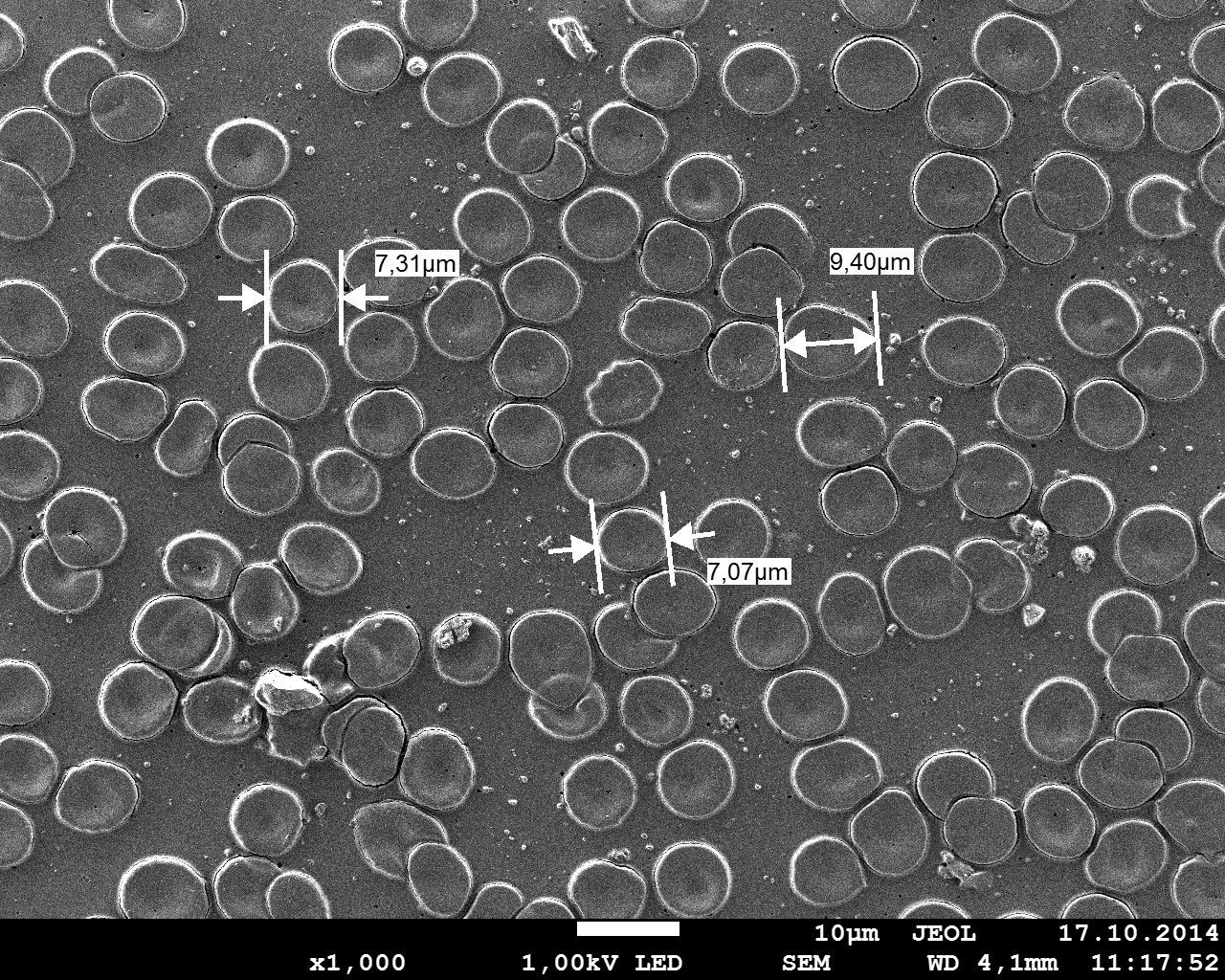
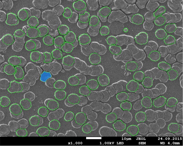
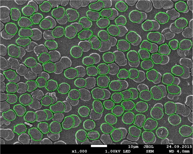
Every year approximately 500,000 women are diagnosed with cervical cancer (WHO, 2018). It is the second most common cancer among women (Ault, 2006) and is very deadly accounting for around 260,000 deaths worldwide (Ault, 2006) and being the leading cause of death by cancer among the female population of developing countries (Denny, 2019).
Mamaeva S.N., Kononova I.V., Ruzhansky M., Nikiforov P.V., Nikolaevа N.A., Pavlov A.N., Fedorova N.F., Huang J., Semenova M.N., Barashkova D.V., Frolova L.S., Maksimov G.V., Using Scanning Electron Microscopy and Atomic Force Microscopy to Study the Formation of Nanoparticles on Red Blood Cell Surface in Cervical Cancer Patients, International Journal of Biomedicine, 10(1): 70-75, 2020. link to the article
Image denoising and segmentation
In some situations, these microscope images may suffer from noise. In our case, “pepper” and “salt” noise and Gaussian noise occur frequently under the high magnification resolution imaging situations. In general, the size of the nanoparticles is around 10-50 pixels in 100,000X, and that of noise can be 3-5 pixels. In this case, it is always difficult to measure the size of nanoparticles with high accuracy. In order to reduce the interference of the noise, we introduce two denoising methods: median filter and L0-norm smoothing filter. The median filter is proved to be helpful to remove “pepper” and “salt” noise, while the L0-norm smoothing algorithm shows a strong ability in filtering out the noise as well as persevering the salient edges.
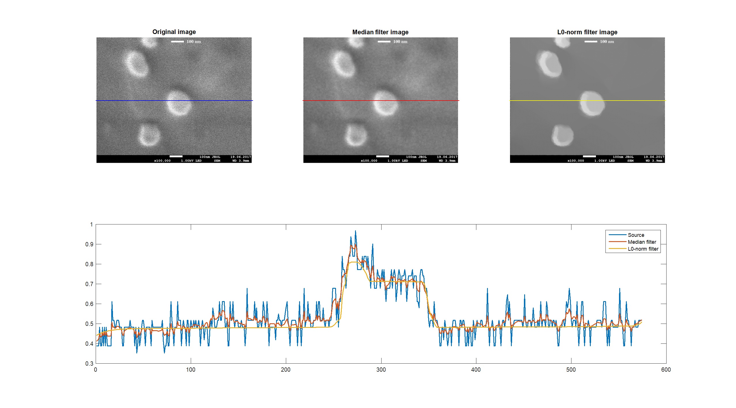
The median filter is very easy to implement. As shown in Figure (in the middle of first row), the “pepper” and “salt” noise are significantly suppressed in comparison of the original image. Moreover, in order to compute the size of nanoparticles more accurately, we introduce the L0-norm smoothing to boost the edges. Mathematically, this method is based on an optimization framework and can preserve the main edges of the nanoparticles while smoothing the local small gradients. The reader is referred to the algorithm (Huang J., Ruzhansky M. et al., 2019) for more details. Obviously, the original data (blue) has strong “pepper” and “salt” noise, and the median filter (red) can reduce some noise but the outline of the nanoparticles is still not clear, while these residual noise can be further reduced with L0-norm filter (yellow).
Some related applications can be seen here, for example, feature extraction algorithms for licence plates based on L0-pseudo-norm sparsity techniques:
Huang J., Ruzhansky M., Feng H., Zheng L., Huang X., Wang H., Feature extraction for license plate location based on L0-norm smoothing. Open Comput. Sci. 2019; 9:28-135. link (open access)
Deep learning for cell analysis
Machine learning (ML) in medical data analysis has drawn great attention in recent years. The rise of deep learning algorithms such as Convolutional Neural Networks (CNN) provides an interesting perspective for automatic medical image analysis. The question is:
“Can ML algorithms improve medical diagnosis?”
The answer is yes, yet, that will be a long journey of scientific exploration …